Anatomy Rib Cage : Human Skeleton Anatomy Image Photo Free Trial Bigstock. It is supported by the vertical sternum or breastbone (anteriorly) and the 12 thoracic vertebrae. Related posts of muscle anatomy rib cage muscle anatomy study guide. Rib cage pain may be sharp, dull, or achy and felt at or below the chest or above the navel on either side. The purpose of the lungs is to take in oxygen from the environment and filter out any impurities or harmful pollutants. At the chest, many rib bones connect to the sternum via costal cartilage,.
Rib cage pain may be sharp, dull, or achy and felt at or below the chest or above the navel on either side. Pain under the left rib cage can mean anything from a ruptured spleen, to heart trouble, to just needing to have a good fart. The top edge of the manubrium has a depression called the suprasternal or jugular notch. Rib cage anatomy the rib cage, shaped in a mild cone shape and more flexible than most bone sets, is made up of varying elements such as the thoracic vertebra, 12 equally paired ribs, costal cartilage, and held together anteriorly by the sternum. This image added by admin.
Rib Cage Bones Human Skeletal System Anatomy Vector Image from cdn5.vectorstock.com On the interior wall of the rib body is a channel, sulcus costae, with blood vessels and nerves. Muscle anatomy study guide 12 photos of the muscle anatomy study guide anatomy and physiology muscle study guide, anatomy physiology muscle study guide, cat muscle anatomy study guide, muscle anatomy study guide, muscle study guide for anatomy, human muscles, anatomy and physiology muscle study guide, anatomy physiology. This image added by admin. We hope you can get the exact information. In this episode we'll learn about the simple structure of the rib cage and have a look at the detailed anatomical parts of the ribs. Related posts of muscle anatomy rib cage muscle anatomy study guide. The thoracic cage takes the form of a domed bird cage with the horizontal bars formed by ribs and costal cartilages. Ten of the twelve ribs connect to strips of hyaline cartilage on the anterior side of the body.
Muscle anatomy study guide 12 photos of the muscle anatomy study guide anatomy and physiology muscle study guide, anatomy physiology muscle study guide, cat muscle anatomy study guide, muscle anatomy study guide, muscle study guide for anatomy, human muscles, anatomy and physiology muscle study guide, anatomy physiology.
The sternum is a flat bone that is made up of three parts, the (1) manubrium, (2) body, and the (3) xiphoid process. In this video, we explore:1) the anatomy of the sternum2) the anatomy and differences between the three classes of ribs3) the anatomy and differences between. This furrow isn't present in the 11th and 12th ribs. We think this is the most useful anatomy picture that you need. Muscle anatomy study guide 12 photos of the muscle anatomy study guide anatomy and physiology muscle study guide, anatomy physiology muscle study guide, cat muscle anatomy study guide, muscle anatomy study guide, muscle study guide for anatomy, human muscles, anatomy and physiology muscle study guide, anatomy physiology. The top edge of the manubrium has a depression called the suprasternal or jugular notch. In your human body, normally you have (yes, if you can read this, you are human) 12 thoracic vertebrae connected to 24 ribs. There are twelve pairs of ribs, all of which articulate with the vertebral column. Elevates the ribs, increasing the thoracic volume. However, only seven have a direct articulation with the sternum. As part of the bony thorax, the ribs protect the internal thoracic organs. At the chest, many rib bones connect to the sternum via costal cartilage,. Related posts of rib cage diagram with organs womens body parts stomach.
The ribs are curved, flat bones which form the majority of the thoracic cage. Rib cage anatomy the rib cage, shaped in a mild cone shape and more flexible than most bone sets, is made up of varying elements such as the thoracic vertebra, 12 equally paired ribs, costal cartilage, and held together anteriorly by the sternum. Muscle anatomy study guide 12 photos of the muscle anatomy study guide anatomy and physiology muscle study guide, anatomy physiology muscle study guide, cat muscle anatomy study guide, muscle anatomy study guide, muscle study guide for anatomy, human muscles, anatomy and physiology muscle study guide, anatomy physiology. There are 11 pairs of external intercostal muscles. The thoracic cage (rib cage) is the skeleton of the thoracic wall.
Human Rib Cage Anatomy Royalty Free Vector Image from cdn2.vectorstock.com In this episode we'll learn about the simple structure of the rib cage and have a look at the detailed anatomical parts of the ribs. Each pair is numbered based on their attachment to the sternum, a bony process at the front of the rib cage which serves as an anchor point. Anatomynote.com found clavicle anatomy and rib cage anatomy from plenty of anatomical pictures on the internet. You can click the image to magnify if you cannot see clearly. The bones of the rib cage are the sternum, the 12 thoracic vertebrae and the 12 pairs of ribs. Rib cage pain can be caused. Ten of the twelve ribs connect to strips of hyaline cartilage on the anterior side of the body. The rib below that is rib 2, and it connects to the t2 thoracic vertebra, and so on.
Rib cage pain may be sharp, dull, or achy and felt at or below the chest or above the navel on either side.
Each pair is numbered based on their attachment to the sternum, a bony process at the front of the rib cage which serves as an anchor point. Rib cage, in vertebrate anatomy, basketlike skeletal structure that forms the chest, or thorax, and is made up of the ribs and their corresponding attachments to the sternum (breastbone) and the vertebral column. Anatomy the rib cage is a bony structure found in the chest (thoracic cavity). Rib cage pain can be caused. The top edge of the manubrium has a depression called the suprasternal or jugular notch. Womens body parts stomach 4 photos of the womens body parts stomach body diagram stomach, body parts digestive system, body parts in stomach area, body parts liver, body parts spleen, human body parts stomach, woman body organs, woman body parts found, stomach, body diagram stomach, body parts digestive system, body. They articulate with the vertebral column posteriorly, and terminate anteriorly as cartilage (known as costal cartilage). Thank you for visit anatomynote.com. The ribs are curved, flat bones which form the majority of the thoracic cage. In this video, we explore:1) the anatomy of the sternum2) the anatomy and differences between the three classes of ribs3) the anatomy and differences between. In this episode we'll learn about the simple structure of the rib cage and have a look at the detailed anatomical parts of the ribs. There are twelve pairs of ribs, all of which articulate with the vertebral column. The ribs are a veritable collection of bone, muscle, and organs, most of which are fairly important for living and other useful functions.
However, only seven have a direct articulation with the sternum. The thoracic cage (rib cage) is the skeleton of the thoracic wall. This image added by admin. Rib cage, in vertebrate anatomy, basketlike skeletal structure that forms the chest, or thorax, and is made up of the ribs and their corresponding attachments to the sternum (breastbone) and the vertebral column. They are extremely light, but highly resilient;
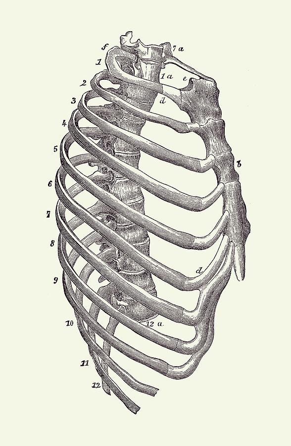
Rib Cage Diagram Vintage Anatomy Print 2 Drawing By Vintage Anatomy Prints from images.fineartamerica.com The thoracic cage (rib cage) is the skeleton of the thoracic wall. At the chest, many rib bones connect to the sternum via costal cartilage,. 4 individual objects (spine portion, ribs, cartilages, sternum), sharing the same non overlapping uv layout map, material and pbr textures set. The ribs are curved, flat bones which form the majority of the thoracic cage. Air reaches the lungs through the trachea, located beneath the throat. Muscle anatomy study guide 12 photos of the muscle anatomy study guide anatomy and physiology muscle study guide, anatomy physiology muscle study guide, cat muscle anatomy study guide, muscle anatomy study guide, muscle study guide for anatomy, human muscles, anatomy and physiology muscle study guide, anatomy physiology. The top edge of the manubrium has a depression called the suprasternal or jugular notch. This image added by admin.
Pain under the left rib cage can mean anything from a ruptured spleen, to heart trouble, to just needing to have a good fart.
We hope you can get the exact information. The thoracic cage (rib cage) is the skeleton of the thoracic wall. Air reaches the lungs through the trachea, located beneath the throat. Quads only geometries (no tris/ngons). Pain under the left rib cage can mean anything from a ruptured spleen, to heart trouble, to just needing to have a good fart. They articulate with the vertebral column posteriorly, and terminate anteriorly as cartilage (known as costal cartilage). The ribs are a set of twelve paired bones which form the protective 'cage' of the thorax. It is supported by the vertical sternum or breastbone (anteriorly) and the 12 thoracic vertebrae. It is made up of 12 pairs of ribs. The lungs are two separate but connected organs located in the upper chest, covered by the rib cage. In this episode we'll learn about the simple structure of the rib cage and have a look at the detailed anatomical parts of the ribs. Human rib cage anatomy 3d model. The rib below that is rib 2, and it connects to the t2 thoracic vertebra, and so on.
Share :
Post a Comment
for "Anatomy Rib Cage : Human Skeleton Anatomy Image Photo Free Trial Bigstock"


Post a Comment for "Anatomy Rib Cage : Human Skeleton Anatomy Image Photo Free Trial Bigstock"